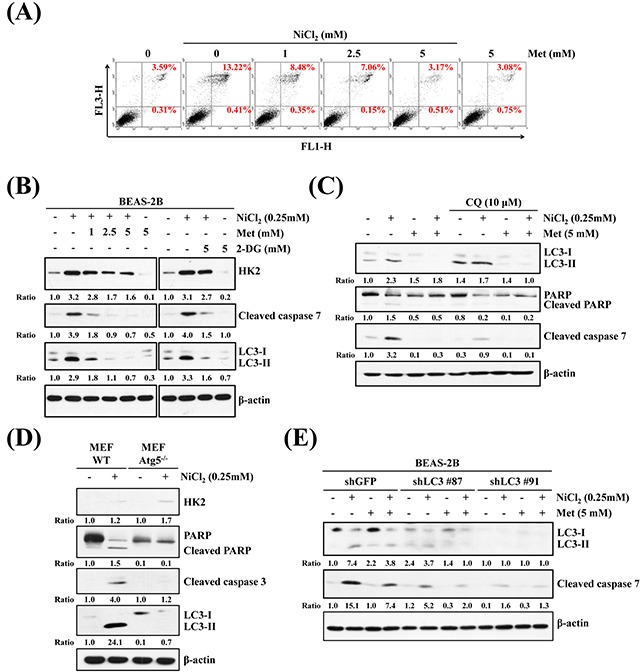
Figure 5. Inhibition of autophagy decreases NiCl2-elicted apoptosis.

(A) Flow cytometry analysis of BEAS-2B cells (1×10⁶ cells/ 6 cm dish) treated with NiCl2 (0, 0.25 mM) and metformin (0, 5 mM) for 48 h, after which cells were stained with Annexin V/PI. (B) BEAS-2B cells (1×10⁶ cells/ 6 cm dish) were treated with NiCl2 (0, 0.25 mM) and metformin (0, 1, 2.5, 5 mM) or 2-DG (0, 5 mM) for 48 h and subjected to western blotting for HK2, cleaved caspase 7 and LC3B. β-actin was used as an internal control. The relative ratios of HK2/β-actin, cleaved caspase 7/β-actin and LC3B-II/LC3-I are shown. (C) Western blot of BEAS-2B cells (1×10⁶ cells/6 cm dish) treated with NiCl2 (0, 0.25 mM) and metformin (0, 5 mM) with or without CQ (0, 10 μM) for 48 h. (D) Atg5 wild type (WT) and Atg5−/− MEF cells (2×10⁵ cells/6 cm dish) were treated with NiCl2 (0, 0.25 mM) for 48 h. β-actin was used as an internal control. The relative ratios of HK2/β-actin, LC3B-II/LC3B-I, cleaved PARP/β-actin and cleaved caspase 3/β-actin are shown. (E) Cleaved caspase 7 and conversions of LC3-I to LC3-II were determined by western blotting after BEAS-2BshLuc and shLC3 cells (1×10⁶ cells/ 6 cm dish) were treated with NiCl2 and metformin for 48 h. β-actin was used as an internal control.
