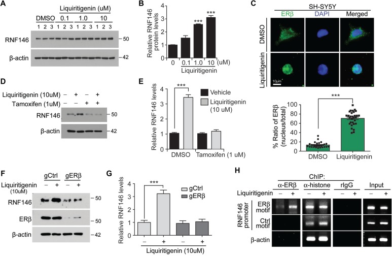
Figure 4. Liquiritigenin induces RNF146 expression via ER activation.
(A) Immunoblot analysis of RNF146 levels in SH-SY5Y cells treated with the indicated concentrations of liquiritigenin for 48 hrs. (B) Quantification of relative RNF146 expression levels (normalized to those of β-actin) from panel A (n = 3). (C) Representative immunofluorescence images showing nuclear translocation of estrogen receptor beta (ERβ) in response to liquiritigenin treatment (37 hrs) in SH-SY5Y cells. Relative distribution of ERβ in the nucleus as normalized to ERβ in the total cell area is shown in the bottom panel (n = 30 cells from two independent experiments). (D) Representative western blots showing that liquiritigenin (10 uM, 48 hrs)-mediated induction of RNF146 expression is blocked by the estrogen receptor antagonist tamoxifen (1 uM, 8 hrs pretreatment). (E) Quantification of relative RNF146 expression levels (normalized to those of β-actin) in the experimental groups in panel C (n = 3). (F) Representative western blots showing that liquiritigenin (10 uM, 48 hrs)-mediated induction of RNF146 expression is blocked by deletion of ERβ by CRISPR-cas9. (G) Quantification of relative RNF146 expression levels (normalized to those of β-actin) in the experimental groups in panel C (n = 3). (H) Chromatin anti-ERβ immunoprecipitation (ChIP) of putative ER responsive element (ERβ motif) within RNF146 promoter region determined by PCR using specific primers. Non ER responsive element within RNF146 promoter (Ctrl motif) and β-actin region were used as negative controls. Immunoprecipitation using either anti-histone antibodies or rabbit IgG was included as ChIP experimental controls. Data are expressed as mean ± SEM. *P < 0.05, **P < 0.01, and ***P < 0.001, unpaired two-tailed student t test or ANOVA test followed by Tukey post hoc analysis.

