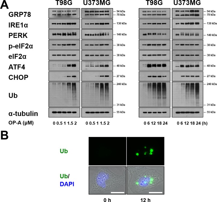
Figure 3. OP-A induces ER stress in glioma cells.
(A) T98G or U373 MG cells were treated with the indicated concentrations of OP-A for 12 h or 2 µM OP-A for the indicated time points and Western blotting of the indicated proteins was performed. α-tubulin was used as a loading control in Western blots. (B) T98G cells treated with 2 µM OP-A for the 12 h were fixed, immunostained using anti-ubiquitin antibody (green), and subjected to immunocytochemistry.

