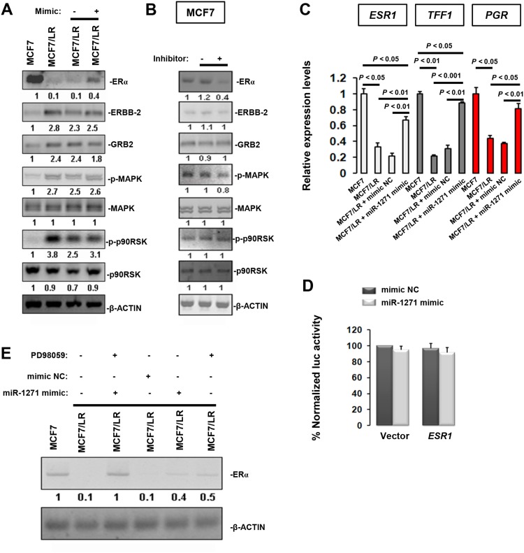
Figure 3. Indirectly modulation of ERα expression by miR-1271.
(A) Immunoblotting analysis of ERα, growth factor receptor ERBB-2, and adapter proteins GRB2 and MAPK pathway in BCa cells. Relative expression levels of target proteins were obtained in each sample by normalization of the expressions of specific targets to that of the β-ACTIN signal. For presentation of data, expression levels of MCF7 were taken as 100% and the others were normalized accordingly, thus allowing semiquantitative comparison. Numbers below the blots represent fold change in protein expression compared with the MCF7 control obtained by densitometric analysis. (B) 48 h after transfection, the untreated MCF7 cells, MCF7 cells transfected with anti-NC and MCF7 cell transfected with anti-miR-1271 cells were subjected to immunoblotting analysis of different targets. (C) 48 h after transfection, untreated MCF7 cells, untreated MCF7/LR cells, MCF7/LR cells transfected with mimic NC and MCF7/LR cells transfected with miR-1271 mimics were subjected to RT-qPCR analysis of different targets. (D) miR-1271 does not target the ESR1 3’UTR in a luciferase reporter assay. (E) Immunoblotting analysis of ERα level in MCF7/LR cells treated with miR-1271 mimic, NC or MAPK pathway inhibitor PD98059.

