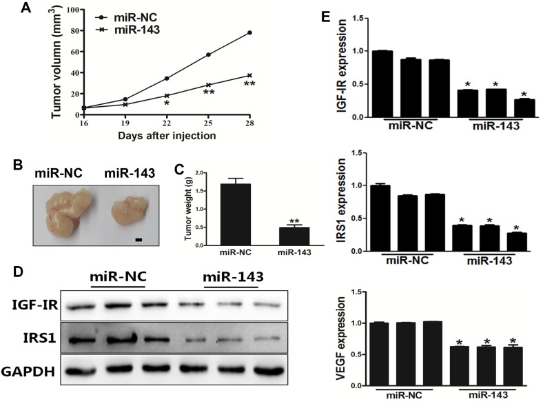
Figure 5. MiR-143 inhibited tumor growth in vivo.
(A) PC-3 cells were transduced with lentivirus carrying miR-143 or miR-NC and selected using puromycin. Cells stably expressing miR-143 or miR-NC at 5x10⁶ were subcutaneously injected into both flanks of male BALB/cA nude mice. Tumor volumes were monitored over time as indicated. (B) After 28 days post implantation, the xenografts were harvested and photographed. Representative pictures from each group were shown. Bar=2 mm. (C) The tumors from each group were weighed and the results showed miR-143 overexpression decreased tumor size and weight in vivo. (D) Total proteins were assayed by Western blotting to determine levels of IGF-IR and IRS1. Levels of GAPDH were used as internal control. (E) Total RNAs were isolated and assayed by qRT-PCR to determine the expression of IGF-IR, IRS1 and VEGF in tumors. Data represent mean±SD. * indicates significant difference at P<0.05; ** indicates significant difference at P<0.01.

