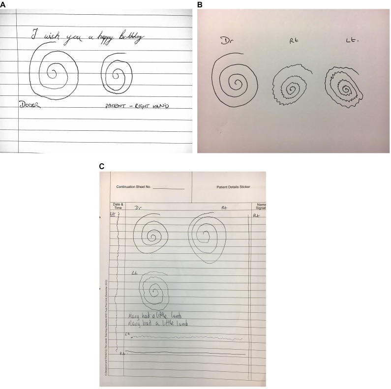
Figure 7.
Parkinson’s disease. (A) The spiral of the patient with Parkinson’s disease (right) is smaller than the clinician’s, with tighter turns. There is no evidence of action tremor. (B) This patient presented with severe bilateral tremor that made it difficult to assess for bradykinesia and rigidity in the upper limbs. It was initially mistaken for essential tremor. There are tightly bunched turns with a unidirectional axis and the patient was also slow to copy the spiral. The oscillations become more widely spaced as the patient speeds up towards the outer sections of the spiral–a pattern frequently seen in Parkinson’s disease, probably reflecting the difficulty with movement initiation. (C) The writing and spiral with the right hand are within normal limits. The left-handed spiral shows a jerky fine amplitude tremor with slight reduction in overall size. The line drawings show a more jerky variation in amplitude and frequency than is usually seen in Parkinson’s disease. This patient has an atypical Parkinson’s action tremor.

