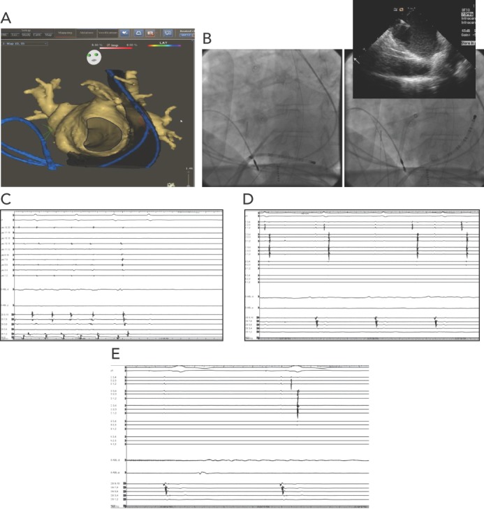
Figure 1: Patient with PLSVC who Underwent AF Catheter Ablation.

A: CARTO map; B: transseptal puncture; C: normal sinus rhythm during isolation of the right pulmonary vein; D: isolation of the PLSVC; E: no reconnection after administration of adenosine. PLSVC = persistent left superior vena cava.
