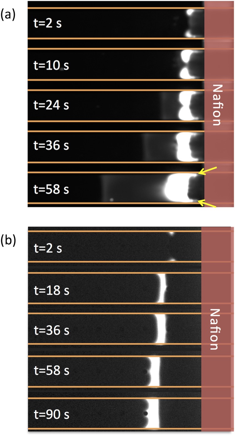
FIG. 2.
Evolution of the focusing band of (a) bovine serum albumin (BSA) protein and (b) a 22mer ssDNA in a nafion-based H-shaped preconcentrator (with only on CEM on one side). The BSA protein and ssDNA were labeled by Alexa Fluor 488 (with different degrees of labeling, the fluorescence intensity are not directly comparable between BSA and ssDNA). The yellow arrows in Fig. 2(a) show the protrusions of the focusing band into the depletion zone near the channel walls. The microchannel is 21μm deep, 200μm wide and 15mm long. The inlet and outlet of the upper channel are biased to 30V and 10V, respectively, while the lower channel is grounded. The background electrolyte is 0.1x phosphate buffered saline (PBS). The detailed experimental setup is described in Ref. 19.

