
Figure 9.
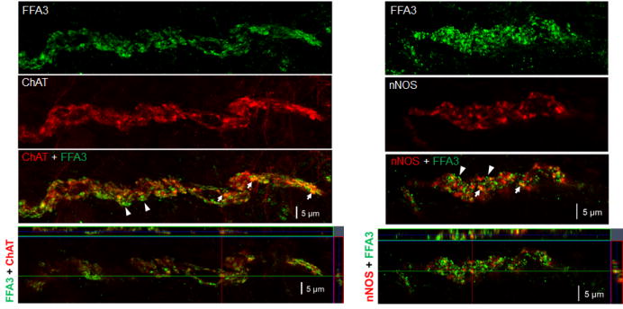
Confocal microscopy with z-stack imaging demonstrating colocalization of FFA3 and cholinergic (ChAT) or nitrergic (nNOS) neural markers in intramuscular nerve fibers in proximal colonic thick sections. The upper three panels depict 3D image whereas the bottom panels depict a confocal image demonstrating the coincidence of green-labeled FFA3 and red-labeled neural marker in varicosities appearing as a yellow color (arrows). Arrowheads indicate lack of colocalization. Bar: 5 μm.