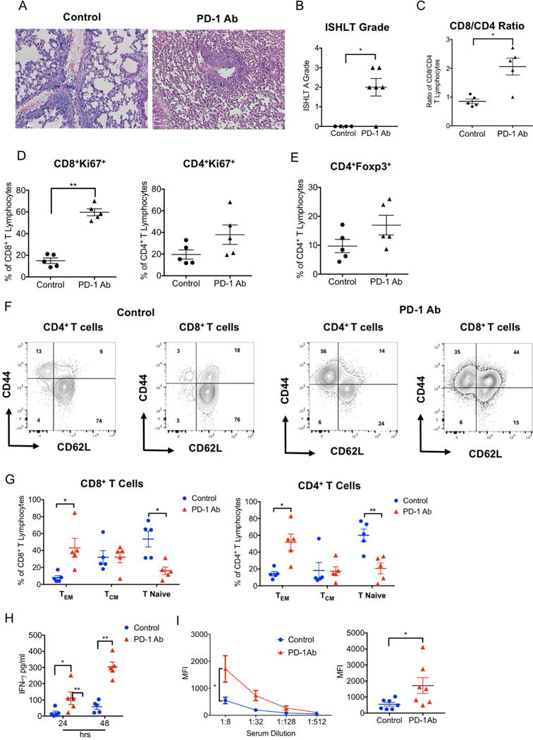
Figure 1. PD-1 blockade prevents costimulation blockade-mediated lung allograft acceptance.

(A) Representative histology, (B) numerical ISHLT rejection grades, (C) intragraft CD8/CD4 T cell ratio, (D) CD8+ and CD4+ T cell proliferative responses and (E) percentage of intragraft CD4+ T cells that express Foxp3 for BALB/c lung allografts seven days after transplantation into control and anti-PD-1-antibody-treated immunosuppressed B6 recipients. Data represent mean ± SEM, *p<0.05, **p<0.01, ***p<0.001 by unpaired T test. (F) Representative contour plot and (G) graphic representation of relative abundance of naïve (CD62LhiCD44low), central memory (CM) (CD62LhiCD44hi) and effector memory (EM) (CD62LlowCD44hi) CD4+ and CD8+ T cells in BALB/c lung allografts seven days after transplantation into control and anti-PD-1-antibody-treated immunosuppressed B6 recipients. (H) IFN-γ levels in supernatants after 24- and 48-hour co-cultures of T cell-depleted Balb/c splenocytes with CD8+ T cells, isolated from Balb/c lung grafts seven days after transplantation into control and anti-PD-1-antibody-treated immunosuppressed B6 recipients. (I) Representative plot (left) and graphic representation (right) of serum IgM alloreactive antibody titers in control and anti-PD-1-antibody-treated immunosuppressed B6 recipients of Balb/c lungs seven days after transplantation. Results are expressed as mean fluorescent intensities (MFI). Data represent mean ± SEM. *p<0.05, **p<0.01 by Mann-Whitney U test.
