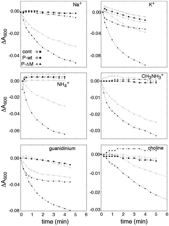
Figure 6.

Inward fluxes in presence of various ions, in cells expressing the Δ-Met FliP variant. Cations are indicated in the figure panels. The counter-ion was Cl- in all cases. Induced cultures contained 10 μM salicylate. Outward water flux was induced by dilution into PBS containing the indicated salts at 0.5 M, as described in Materials and Methods. Measurements began a few (6-8) seconds after mixing, and monitored the slower return toward the initial absorbance level. Experiments were carried out at room temperature (ca. 20° C). Open symbols indicate non-induced cultures, and filled symbols cultures with FliP expression induced with 10 μM salicylate; ‘cont’ indicates the vector-only controls not expressing FliP.
