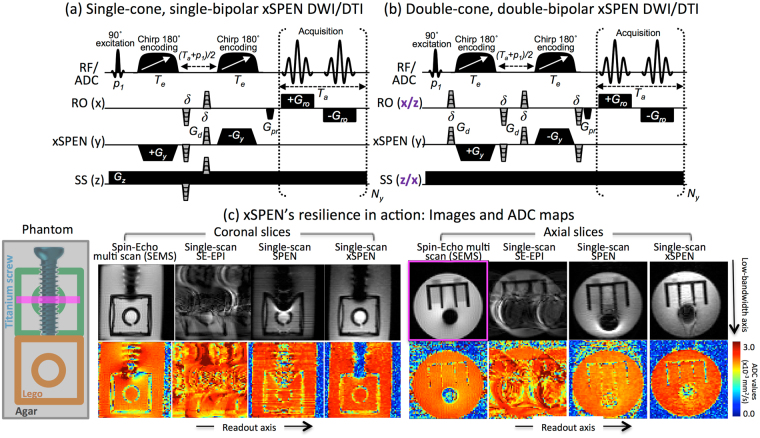
Figure 1.
(a,b) xSPEN diffusion pulse sequences assessed in this study, involving in all cases a slice-selective 900 excitation pulse (p1) followed by two 1800 chirped pulses. In (a), a single diffusion-weighting PGSE block is placed in the pre-encoding (T a + p 1)/2 delay required for targeting the desired FOV under full-refocusing. In (b) two PGSE diffusion blocks are placed on both sides of the 1800 chirped pulses to enable a larger diffusion weighting, and both RO and SS axes are alternated among x and z orientations in order to overcome the otherwise dominating b zz-weighting derived from the G z gradient. The RF/ADC line displays the pulses and signal acquisition; RO, SPEN and SS display orthogonal gradient directions; G d are diffusion-weighting gradients of duration δ and stepped amplitudes and/or different directions (in grey); G pr, purge gradients; G ro, readout acquisition gradients; T a, acquisition time. (c) MRI results obtained on a preclinical 7 T scanner for an agar phantom containing a titanium screw and two Lego pieces arranged as indicated on the left, imaged by a reference multi-shot spin echo (SEMS) and by several (SE-EPI, SPEN, xSPEN) single shot methods. In the latter case, the sequence in panel (a) was used with G d = 0 (so-called b o images). Indicated in magenta is the position of the axial slice within the coronal rendering; the coronal slices were not wide enough to capture the back (green) Lego piece. Notice that while metal-induced field distortions are evident in all four methods, the xSPEN results most faithful reproduce the original phantom distribution and provide the most reliable ADC map.

