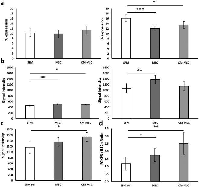
Figure 4.
Increased immunosuppressive/anti-inflammatory T cell subsets following CM-MSC treatment at 3 days post induction of arthritis. (a) Surface staining of CD4 assessed proportion of CD4+ cells in spleens (left) and lymph nodes (right). (b) Intracellular staining for key cytokines characteristic of Treg (FOXP3) in spleens (left) and lymph nodes (right) and (c) Th2 (IL4) following 4 hours culture with inhibition of protein transport using Brefeldin A for T cells from CM-MSC and MSC treated mice and SFM treated controls (mean fluorescence intensity (MFI)). (d) The ratio of percentage FOXP3+ to IL17a+ cells (Treg:Th17) was calculated showing an improved ratio of FOXP3:IL17a expression following CM-MSC co-culture. All data were obtained from AIA mice at day 3 post arthritis induction. (*p < 0.05; **p < 0.01; ***p < 0.001).

