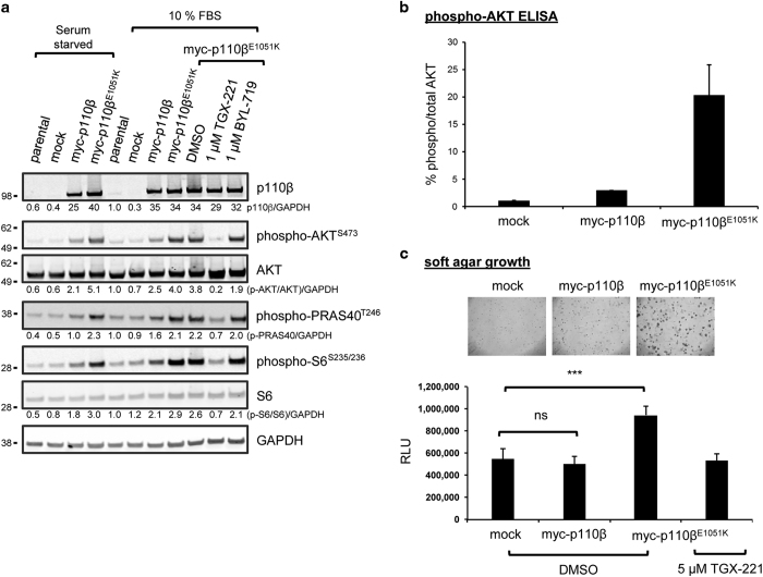
Figure 2.
Exogenous expression of p110βE1051K promotes PI3K pathway activation and anchorage-independent growth. (a) Rat2 cells stably expressing myc-p110β or myc-p110βE1051K or cells transduced with empty vector (mock) were maintained in growth conditions (10% FBS) or serum starved in 0.5% FBS overnight prior to lysis. Lysates of cells were separated by SDS-PAGE and probed with antibodies to detect p110β expression, phosphorylated proteins, or GAPDH loading control as indicated. Fluorescent intensity of bands was quantified and normalized to GAPDH and then to signals from parental Rat2 cell lysates and is shown below blots to 2 sig. fig. (b) The level of AKTS473 phosphorylation in transduced Rat2 cell lysates was determined using a multiplexed phospho-/total electrochemiluminescent ELISA assay. Data are the mean±s.e.m. of duplicate samples from two independent experiments. (c) Soft agar growth assay to compare the effect of stable expression of myc-p110β and myc-p110βE1051K on the growth of Rat2 cells in soft agar with control cells harboring empty vector (mock). Rat2 cells were plated in soft agar, and growth was imaged after 14 days by light microscopy and quantified with Alamar Blue viability assay. Data are the mean±s.e.m. of duplicate samples from two independent experiments. On one test occasion, Rat2 cells expressing myc-p110βE1051K were also cultured in the presence of 5 μM TGX-221. ***indicates P=0.0005; ns, not significant.

