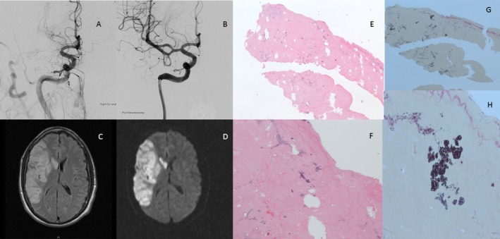
Figure 2.

(A) Cerebral digital subtraction angiography (DSA) showing right M1 occlusion. (B) DSA after endovascular thrombectomy with aspiration through an intermediate catheter. Complete right middle cerebral artery (MCA), infarction post‐EVT on (C) T2‐weighted‐fluid‐attenuated inversion recovery (T2‐FLAIR), and (D) diffusion‐weighted imaging (DWI). Hematoxylin and eosin (H&E) stains at (E) low (20X) and (F) high magnification (40X), respectively. Gram stain of the clot showing gram‐positive cocci consistent with septic embolus at (G) low (20X) and (H) high magnification (40X), respectively.
