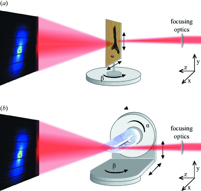
Figure 1.
(a) Three-dimensional scanning SAXS setup to measure thinly sliced samples. The sample is scanned through the focused beam in x and y at different rotation angles β around y. (b) SAXS tensor tomography setup to measure three-dimensional samples. The sample is scanned through the focused beam in x and y at n orientations which are described by the rotation matrix  .
.

