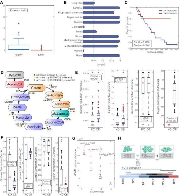
A boxplot describing the expression of FUT9 in tumor vs. healthy colon tissues.
Q‐value for CN of FUT9 in 12 different cancer types, the dashed line represents a significance threshold of 0.25.
Kaplan–Meier survival curve for FUT9 expression (top and bottom 0.5 quartiles).
The TCA cycle and its associated enzymes that are increased in stage 4 colorectal cancer (red), predicted to increase following FUT9 KD (yellow) and increase following FUT9 KD experimentally (green).
Boxplot showing the distribution of biomass production, glucose consumption, lactate production, and oxygen consumption in adenoma state when FUT9 is knocked down (KD) and overexpressed (OE).
Boxplot showing the distribution of biomass production, glucose consumption, lactate production, and oxygen consumption in cancer state when FUT9 is knocked down (KD) and overexpressed (OE).
Boxplots sowing the MOMA scores obtained by the knockdown of FUT9 in stages 1–4.
Upper panel: Colorectal adenoma–carcinoma sequence. Middle panel: the emerging role of FUT9 in colorectal tumor progression. Lower panel: Correlation heat map of FUT9 copy number (CN) and early‐ and late‐stage prognostic markers of colorectal cancer.
Data information: (A, E–G) On each box, the central mark indicates the median, and the bottom and top edges of the box indicate the 25
th and 75
th percentiles, respectively. The
P‐values are for one‐sided Wilcoxon rank‐sum test.

