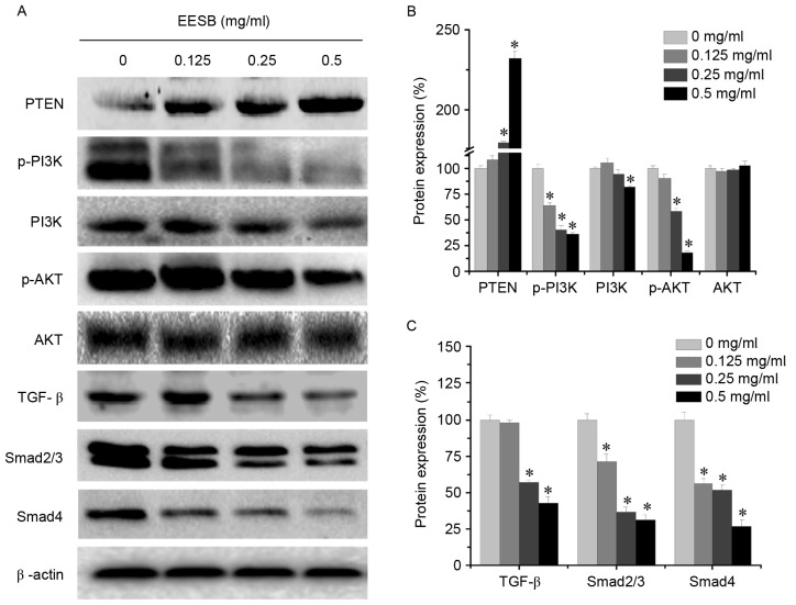
Figure 5.
Effect of EESB on the activation of PI3K/AKT and TGF-β/Smad signaling pathways. Cells were treated with EESB at different concentrations for 24 h. (A) PI3K/AKT and TGF-β/Smad protein expression levels were determined by western blotting. β-actin was used as the internal control. Images are representatives of three independent experiments. Densitometric analysis for (B) PTEN, p-PI3K, PI3K, p-AKT and AKT and (C) TGF-β, Smad2/3 and Smad4. Data are expressed as the mean ± standard deviation and were normalized to the mean protein expression of the untreated control (100%). *P<0.05. vs. the control cells. EESB, ethanol extract of Scutellaria barbata D. Don; PI3K, phosphoinositide 3-kinase; TGF-β, transforming growth factor-β; PTEN, phosphatase and tensin homolog; p, phospho.

