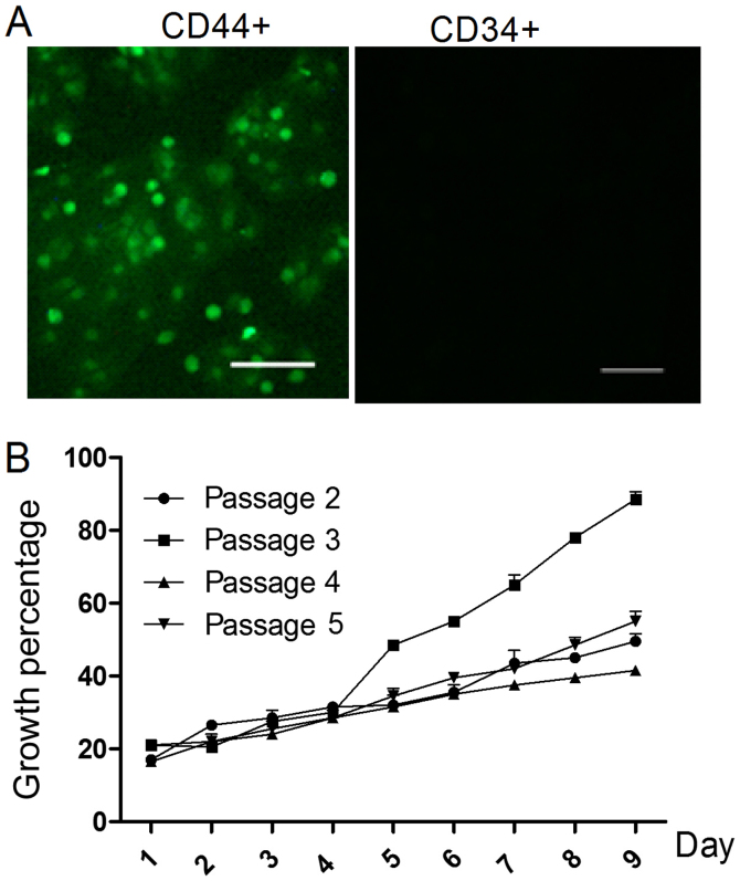
Figure 1.
Identification of rMSCs. (A) Immunofluorescence of cell markers, CD44 and CD34, in cultured rMSCs. Scale bar, 25 µm. (B) Cellular proliferation during the second to fifth cell passages (as determined at an absorbance of 570 nm). CD, cluster of differentiation; rMSCs, rabbit mesenchymal stem cells.

