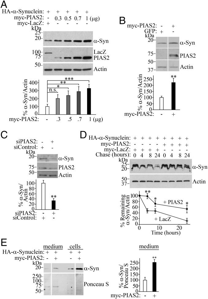
Fig. 3.
SUMOylation by PIAS2 regulates α-synuclein levels and degradation. (A) PIAS2 increases the steady-state levels of α-synuclein in transfected HEK293 cells. Graph depicts the percentage of α-synuclein steady-state levels relative to β-actin at increasing amounts of myc-PIAS2. (B) Endogenous levels of α-synuclein increase upon myc-PIAS2 transfection. (C) PIAS2 knockdown decreases endogenous α-synuclein levels. HEK293 cells were transfected with siRNA control or siRNA to PIAS2. Endogenous levels of α-synuclein were determined using anti–α-synuclein antibody. (D) PIAS2 prolongs α-synuclein half-life in transfected HEK293 cells treated with 50 μM cycloheximide. Graph depicts α-synuclein levels relative to β-actin in the presence of PIAS2 (●) or control LacZ (○). (E) PIAS2 elevates α-synuclein in extracellular medium. Levels of α-synuclein in intracellular and extracellular HEK293 cell compartments were monitored with anti-HA (Upper). Total protein loading was determined by Ponceau S staining (Lower). Graph depicts the percentage of α-synuclein in the medium relative to total protein loading monitored by Ponceau S staining. Values represent the average ± SEM of three to four experiments. Different from control at *P < 0.05, **P < 0.01, and ***P < 0.001. Repeated-measures one-way ANOVA with Bonferroni post hoc test (A) or Student’s t test (B–E). n.s., not significant.

