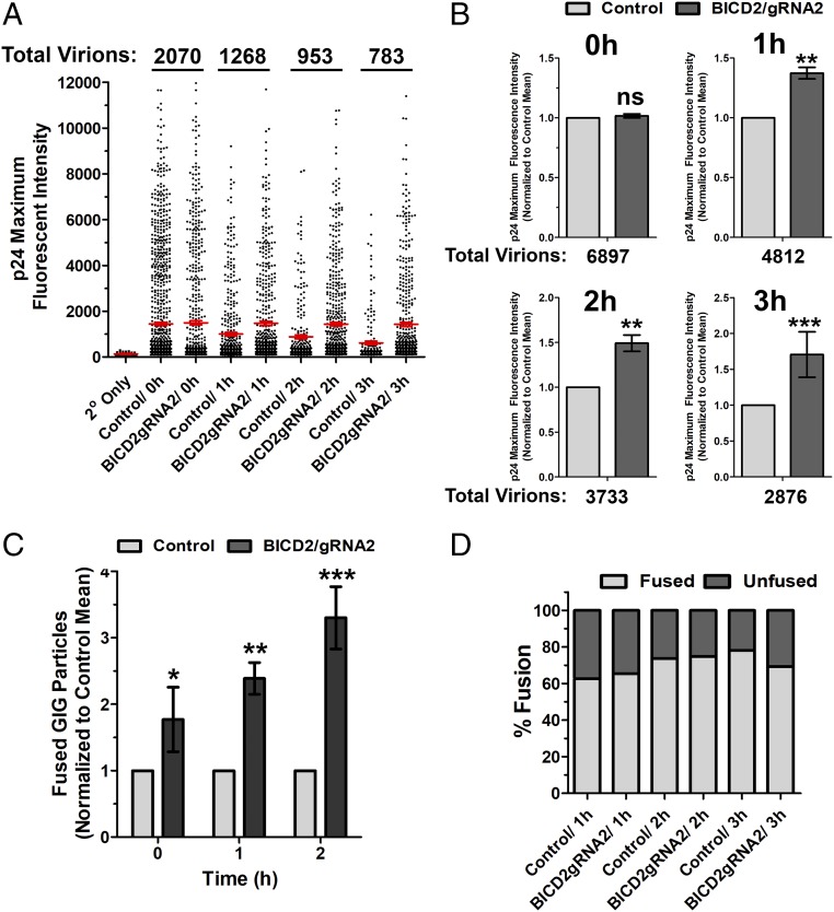
Fig. 3.
BICD2 depletion delays HIV-1 uncoating as measured using an in situ uncoating assay. Control and BICD2-depleted HeLa TZM-bl cells were synchronously infected with S15-mCherry/GIG JRFLg-HIV-1. (A) Cells were fixed at the indicated time points, and the p24 intensities associated with individual virions lacking the S15 membrane label (1–3 h postinfection) or all virions (0-h point) are shown. Red line represents average p24 intensity measured for all fused viruses at the indicated time points; 20–25 cells were imaged at each time point. Error bar represents SEM. (B) Data from three independent experiments, as shown in A, were normalized to the mean p24 intensity observed in control cells and averaged. (C) Fused GIG particles in control and BICD2-depleted cells from three independent experiments. Normalized to the number of cells observed in control cells at the indicated time points. (D) Percent fusion was calculated from the experiment described in A. Data are representative of three independent experiments. ***P < 0.001, **P < 0.01, *P < 0.05; ns, not significant.

