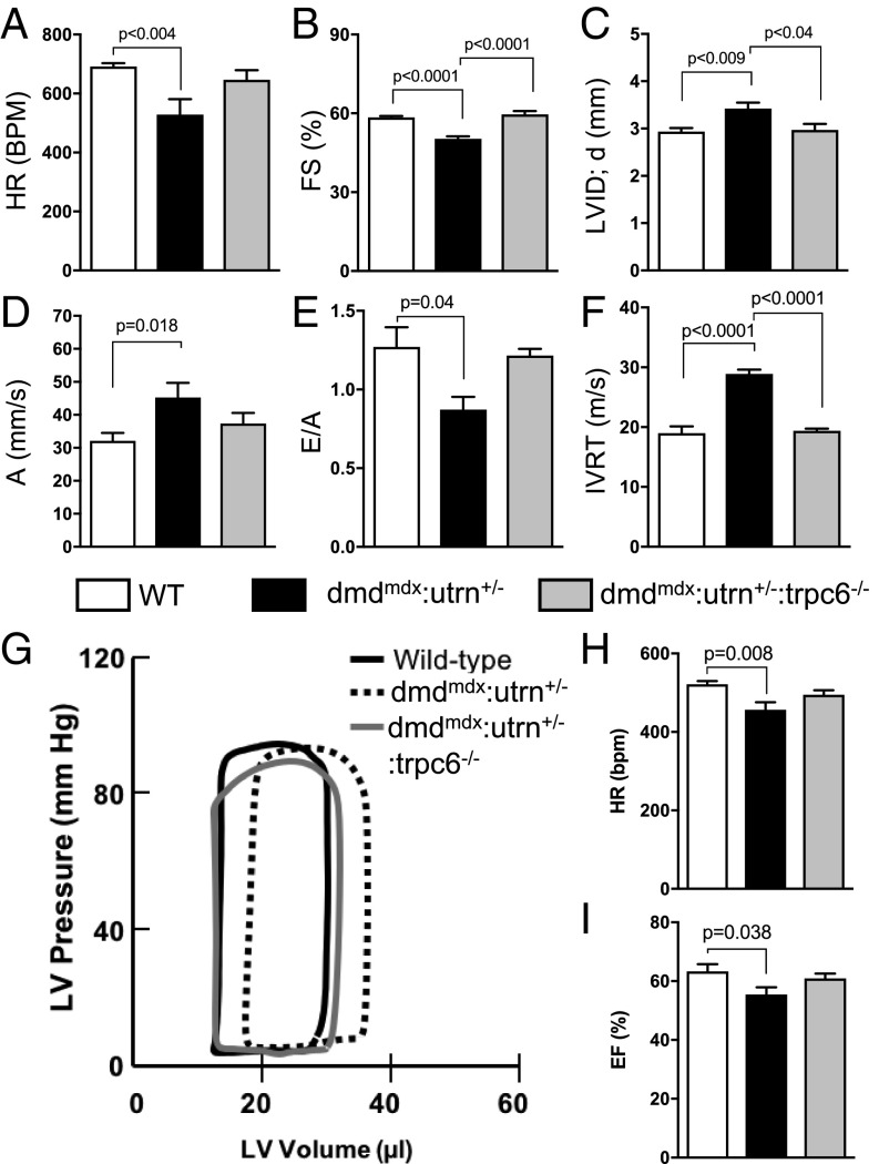
Fig. 4.
LV dilatation increases preload to depress dmdmdx:utrn+/− cardiac function. (A–F) In vivo cardiac function assessed by echocardiography. A, atrial contraction tissue velocity; BPM, beats per minute; E/A, early-to-atrial filling ratio; FS, fractional shortening; HR, heart rate; IVRT, isovolumic ventricular relaxation time; LVID;d, LV diastolic dimension. Pathological changes in all parameters were restored with gene deletion of Trpc6. (WT, n = 8; dmdmdx:utrn+/− dystrophy model, n = 6; and dmdmdx:utrn+/−:trpc6−/− dystrophy model lacking Trpc6, n = 4.) (G–I) Representative pressure-volume loop traces and summary data at rest. The HR and ejection fraction (EF) were reduced in dmdmdx:utrn+/− and restored by Trpc6 deletion. (n = 13, n = 19, and n = 17 for the three groups, respectively.)

