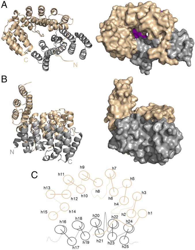
Fig. 1.
Structure and overview of CpcE/F heterodimer. Subunits E (brown) and F (gray) correspond to chains A and B in PDB ID code 5N3U. In the asymmetric unit, there is one heterodimer consisting of two chains containing 15 (CpcE) and 10 (CpcF) helices. Both subunits appear as α-solenoid proteins in the shape of twisted crescents. Each is composed of two layers of α-helices, and the two subunits are arranged in a spooning fashion (see main text for details). (A) Top view into the cavity formed between subunits E and F that fits a PCB chromophore. The cavity has a wide opening at the top and a smaller one at the bottom that is closed by three loops and two short helices that are in part visible (magenta) looking down the cavity (upper right). (B) Side view with subunit F in front, and the handle formed by the C-terminal helices of subunit E on Left. (C) Schematic structure, seen from the top into the cavity as in A. Dashed lines indicate unresolved stretches; curved lines indicate extended structures.

