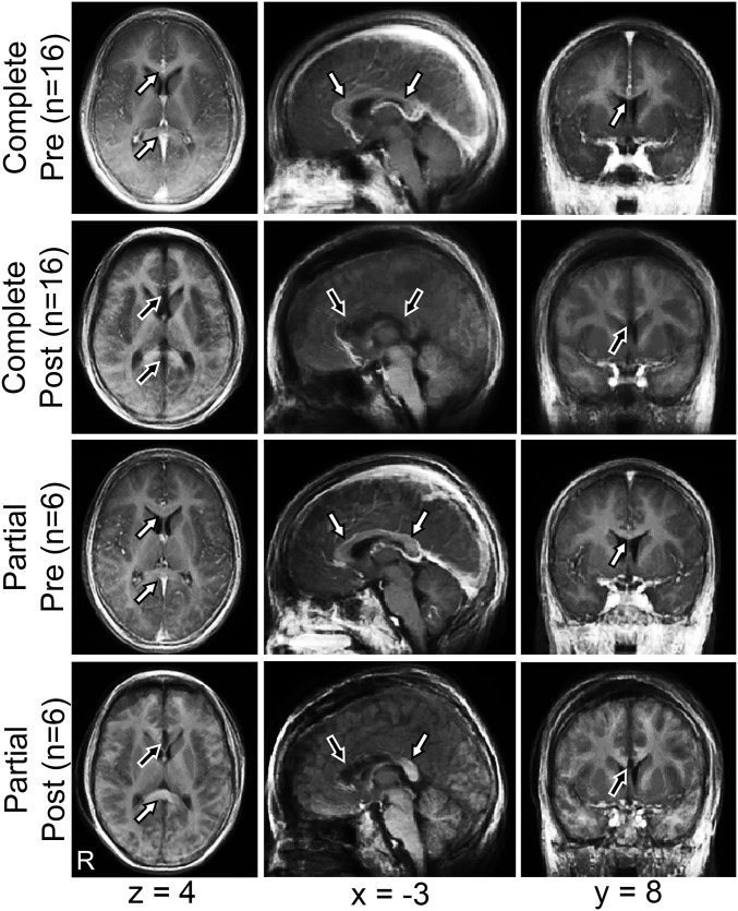
Fig. 1.
Anatomic imaging precallosotomy and postcallosotomy. Mean T1-weighted images before (precallosotomy) and after (postcallosotomy) complete and partial callosotomy, represented in atlas space (right hemisphere on Left). MNI152 coordinates of axial, sagittal, and coronal planes are listed. White arrows indicate intact CC, and black arrows indicate areas of divided CC. Note residual splenium after partial callosotomy.

