Fig. 2.
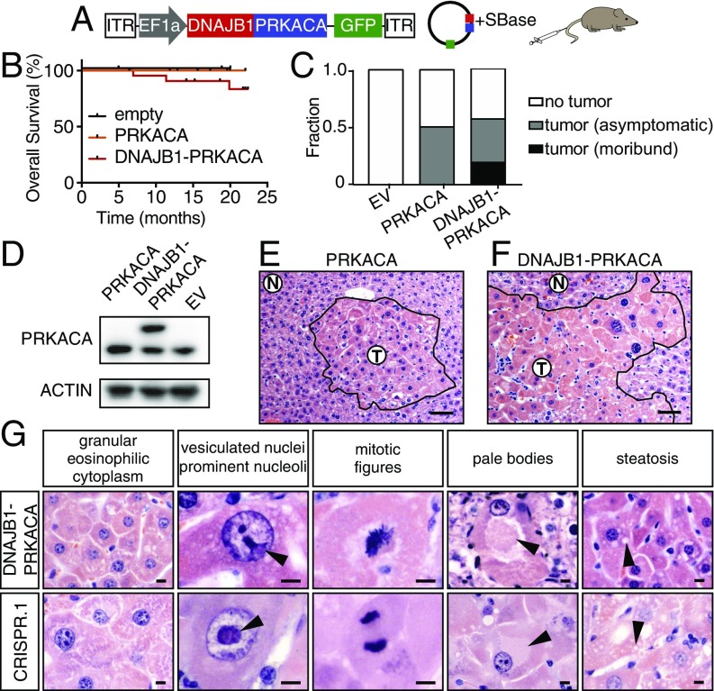
DNAJB1–PRKACA fusion drives liver tumorigenesis. (A) Schematic of the Sleeping Beauty transposon strategy to deliver DNAJB1–PRKACA fusion cDNA to young adult livers (ITR, inverted terminal repeat). (B) Overall survival of mice injected with cDNA encoding DNAJB1–PRKACA (n = 23), wild-type PRKACA (n = 12), or empty vector (n = 4). (C) Fraction of mice harvested with no detectable tumor, asymptomatic mice with histologically detectable disease, or moribund mice with tumors expressing empty vector (EV), wild-type PRKACA, or DNAJB1–PRKACA fusion. (D) Western blot of liver progenitor cells 4 d after viral transduction with the indicated cDNA in vitro. (E) Cluster of atypical hepatocytes in liver injected with wild-type pT3-PRKACA. (Scale bar, 50 μm.) (F) Tumor generated by pT3-DNAJB1–PRKACA. (Scale bar, 50 μm.) (G) Higher-magnification images highlighting common murine FL-HCC features: large granular, eosinophilic cytoplasm; vesiculated nuclei with prominent nucleoli (arrowheads); mitotic figures; pale bodies; and steatosis. (Scale bars, 10 μm.)

