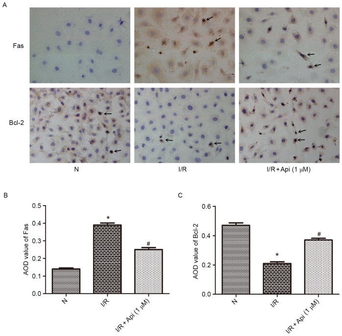
Figure 7.
Immunohistochemical analysis examining the expression levels of Fas and Bcl-2 in NRK-52E cells. (A) Fas and Bcl-2 expression levels analyzed by immunohistochemical staining (original magnification, ×400). The AOD values of (B) Fas and (C) Bcl-2 were measured. Bars represent the means ± standard deviation (n=6). *P<0.01 vs. normal group; #P<0.05 vs. I/R group. I/R, ischemia-reperfusion; N, normal; api, apigenin treatment; AOD, average optical density; Bcl-2, B-cell lymphoma 2.

