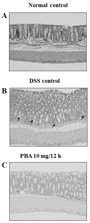
Figure 5.

Histopathology of colon tissue. (A) Low-power view of a longitudinal section of a normal colonic wall. Note the crypts with abundant goblet cells. (B) Chronic inflammation in the lamina propria of mice with experimental colitis. Note the loss of goblet cells, the frequently enlarged nuclei of the absorptive cells and the unequivocal mucosal erosion because of the loss of surface epithelia. Loss of goblet cells (indicated by black arrows). (C) No signs of inflammation in the colonic wall of PBA mice. Magnification, ×200. DSS, dextran sulfate sodium; PBA, sodium 4-phenylbutyrate.
