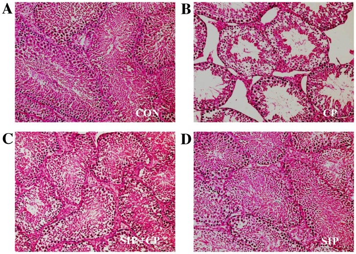
Figure 1.
Histopathological observation of testicular seminiferous tubules in (A) control, (B) cp-treated, (C) SIP+CP treated and (D) SIP-treated mice.. Testicular seminiferous tubules were observed by optical microscopy and hematoxylin and eosin staining techniques. Images were captured at a magnification of ×200. CON, control mice; CP, CP-treated mice; SIP+CP, mice treated with squid ink polysaccharide and CP; SIP, squid ink polysaccharide-treated mice. Scale bar 0.05 mm.

