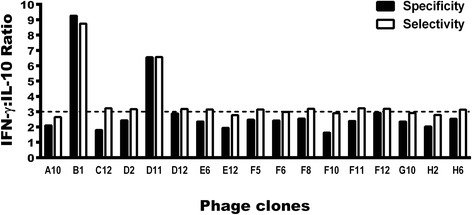
Fig. 1.

Evaluation of selectivity and specificity of the phage clones. PBMCs were purified from blood samples from asymptomatic and symptomatic VL patients and non-infected subjects. Cells were non-stimulated (medium) or stimulated with each clone, as well as by Wild-type (WT) and Random clones (1010 phages each), for 48 h at 37 °C in 5% CO2. IFN-γ and IL-10 levels were measured in the culture supernatants by an ELISA capture. Black bars indicate the specificity of the clones, which was calculated by dividing the IFN-γ and IL-10 values obtained of each evaluated clone through respective values of these cytokines obtained after the WT phage stimulus, using PBMCs from healthy subjects. With the corrected values, the ratio between the IFN-γ and IL-10 levels with these new results was calculated, and the specificity of the each clone was defined and is shown here. White bars indicate the selectivity, which was calculated by dividing the IFN-γ and IL-10 values obtained of each evaluated clone through respective values of these cytokines obtained after the Random phage stimulus, using PBMCs from VL patients. With the corrected values, a ratio between the IFN-γ and IL-10 levels with these new results was calculated, and the selectivity of each clone was defined and is shown here
