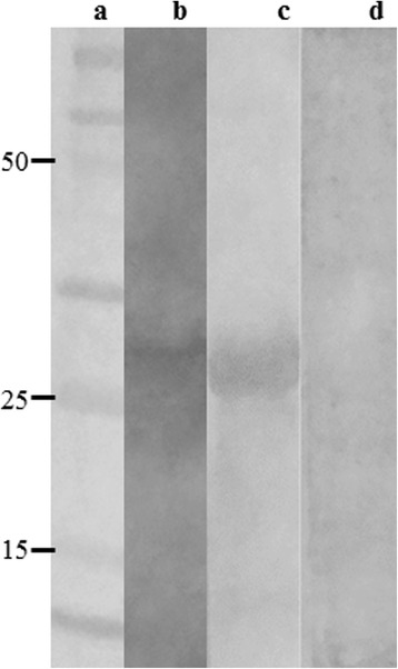
Fig. 2.

Immunoblotting assays. A molecular weight marker (Lane a), 20 μg L. infantum SLA (Lane b) and 10 μg rLiHyp (Lanes c, d) were electrophoresed on a 20% SDS-PAGE and blotted onto nitrocellulose membranes. Blots were incubated with pools of sera from B1 phage-vaccinated mice (Lanes b and c) or from naive mice (lane d), and were revealed by adding chloronaphtol, diaminobenzidine, and H2O2. A scan from the blots is shown here. The black arrow indicates the rLiHyp protein (~28.0 kDa)
