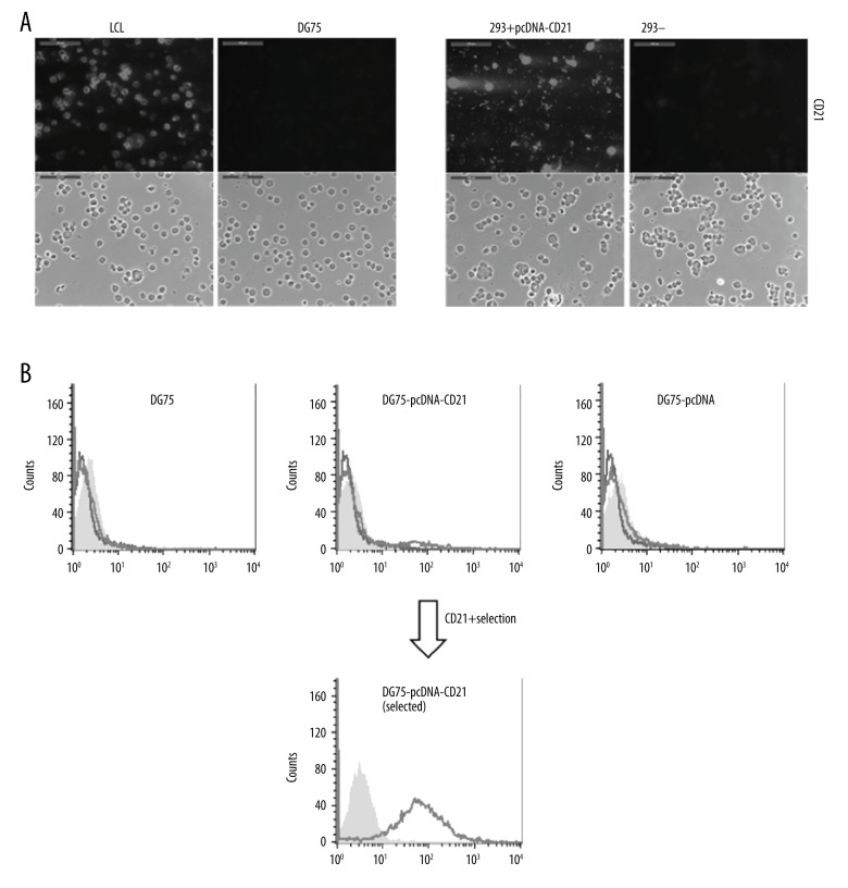
Figure 2.
Expression of CD21 on DG75 cells using generated pcDNA3.1(+)-CD21. (A) The generated CD21 expression plasmid (DG75-pcDNA3.1(+)-CD21) was tested by transient expression of 293 cells for 3 d, which was confirmed by immunostaining. LCLs and DG75 cells were used as positive and negative controls for CD21 expression, respectively. Untransfected 293 cells were also compared in parallel as a negative control. Both immunofluorescent and phase-contrast pictures were taken, and scale bars represent 100 μm. (B) Stable transfection of DG75 cells with pcDNA3.1(+)-CD21 plasmid was performed by electroporation. FACS analysis of untransfected cells (DG75) and cells transfected with pcDNA3.1(+)-CD21 plasmid (DG75-pcDNA3.1(+)-CD21) or empty vector (DG75-pcDNA3.1(+)), the expression of CD21 was tested after establishment of the stable cell line, and the selected CD21+ DG75 was confirmed by FACS analysis. The grey shading represents unstained samples alone, the black line represents samples stained with secondary fluorescent antibody alone, and the light dark line represents samples stained with anti-CD21 antibody coupled with secondary fluorescent antibody. pcDNA3.1(+) is shortened to pcDNA for convenience.

