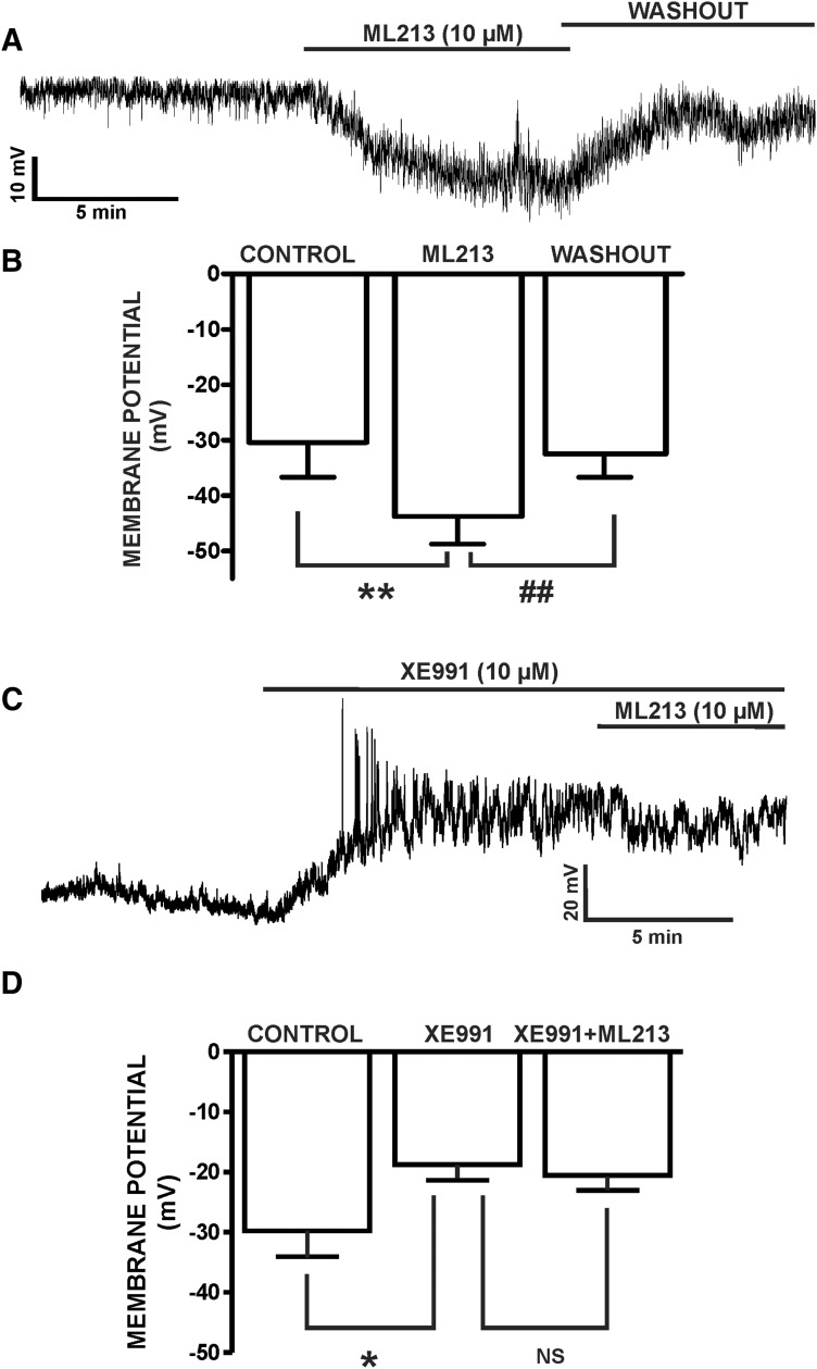
Fig. 10.
ML213 hyperpolarizes the resting membrane potential in freshly isolated DSM cells. (A) A representative membrane potential recording in current-clamp mode illustrating that ML213 (10 µM) hyperpolarized the DSM cell membrane potential in a freshly isolated DSM cell and that this effect was reversible by washout. (B) Summarized data for the hyperpolarizing effects of ML213 (10 µM) on the DSM cell membrane potential (n = 5, N = 5; **P < 0.01) and for the washout (n = 5, N = 5, ##P < 0.01). (C) A representative membrane potential recording in current-clamp mode demonstrating that XE991 (10 µM) depolarized the DSM cell membrane potential and blocked ML213 (10 µM) hyperpolarization in a freshly isolated DSM cell. (D) Summarized data for XE991-induced (10 µM) depolarization in freshly isolated DSM cells and the lack of ML213 (10 µM) hyperpolarizing effects in the presence of 10 µM XE991 (n = 8, N = 4; *P < 0.05; two-tailed paired Student’s t-test), NS, nonsignificant. See Supplemental Fig. 10 for display of summary data as scatterplots with means.

