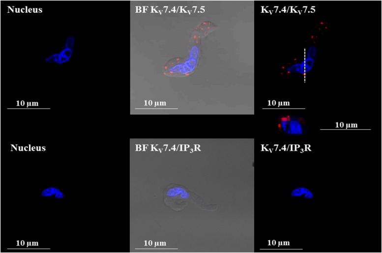
Fig. 12.
PLA shows that KV7.4/KV7.5 heteromeric channels are expressed in isolated guinea pig DSM cells. Images of a freshly isolated DSM cell stained with the combination of anti-KV7.4 and anti-KV7.5 channel antibodies (top panels). PLA fluorescent signals are shown in red. No PLA fluorescent signals were observed when the anti-KV7.4 antibody was coincubated with the anti-IP3R antibody (bottom panels). An orthogonal cross-sectional image of the DSM cell displaying PLA signals was obtained after z-stack scans collected along the viewing plane indicated by the dashed (- - -) lines and is represented directly below the top right panel. BF, bright field; DAPI, 4′,6-diamidine-2′-phenylindole dihydrochloride (nuclear staining shown in blue).

