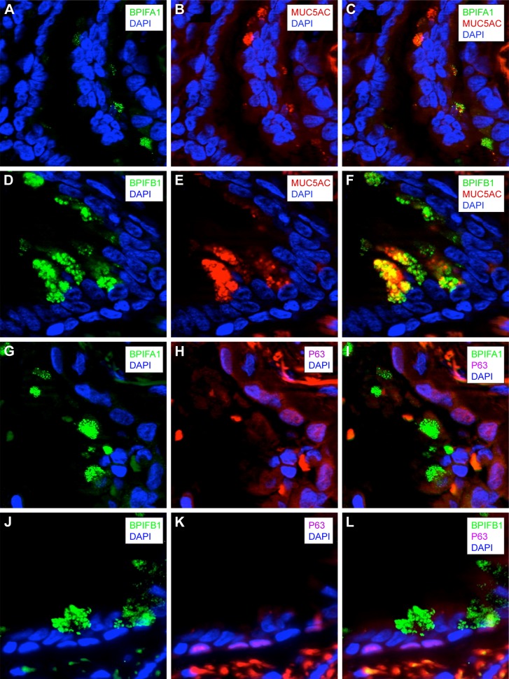
Figure 6.
Localization of BPIFA1 and BPIFB1 expression in lung tissue by double immunofluorescence staining.
Notes: Immunofluorescence staining was performed on paraffin-embedded lung tissue of a COPD GOLD II patient for BPIFA1, BPIFB1, P63, and MUC5AC. Blue DAPI staining is used to stain the cell nucleus. Immunofluorescence staining of BPIFA1 and DAPI (A), MUC5AC and DAPI (B), the merge of the two previous stainings (C). Immunofluorescence staining of BPIFB1 and DAPI (D), MUC5AC and DAPI (E), the merge of the two previous stainings (F). BPIFB1 shows strong colocalization with MUC5AC, whereas colocalization of BPIFA1 and MUC5AC is limited. Immunofluorescence staining of BPIFA1 and DAPI (G), P63 and DAPI (H), the merge of the two previous stainings (I). Immunofluorescence staining of BPIFB1 and DAPI (J), P63 and DAPI (K), the merge of the two previous stainings (L). BPIFA1 and BPIFB1 are not localized in basal cells.
Abbreviations: BPIF protein, bactericidal/permeability-increasing fold-containing protein; COPD, chronic obstructive pulmonary disease; GOLD, Global Initiative for Chronic Obstructive Lung Disease.

