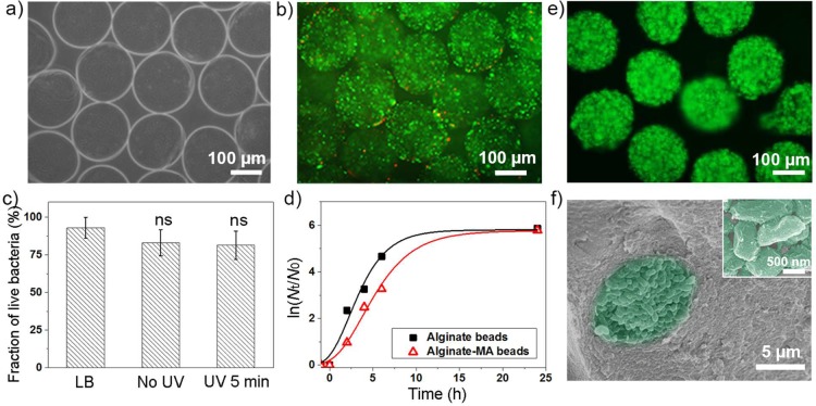
Figure 6.
(a) Optical microscopy image of bacteria loaded alginate-MA beads. (b) Live/dead staining of bacteria inside beads after 5 min UV irradiation, 0.1 wt % Irgacure 2959; green staining indicates viable bacteria, red staining indicates dead bacteria. (c) Fraction of live E. coli pLuxR-GFP before encapsulation, after encapsulation without UV irradiation, and after 5 min UV irradiation; the data are presented as arithmetic mean ± standard deviation calculated from at least three biological replicates, one-way ANOVA. (d) Natural logarithm plot of normalized E. coli pLuxR-GFP population density inside alginate and alginate-MA beads incubated in LB broth. (e) FDA staining of live bacteria entrapped in alginate-MA beads after 24 h incubation in LB media at 37 °C. (f) SEM image of E. coli pLuxR-GFP cluster inside alginate-MA bead; inset is a high-magnification image of entrapped E. coli pLuxR-GFP.

