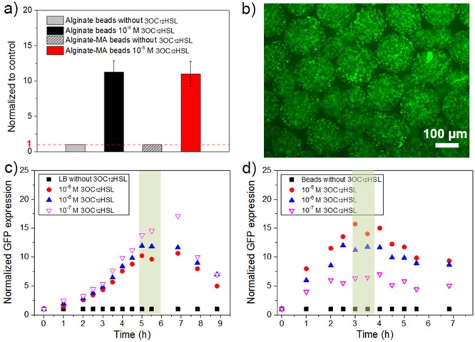
Figure 9.
(a) Fluorescence intensity (normalized to that of alginate-based beads in the absence of autoinducers) of E. coli pLuxR-GFP in alginate-based beads exposed to 1.0 × 10⁻⁶ mol/L 3OC12HSL. (b) Fluorescence microscopy image of beads loaded with E. coli pLuxR-GFP in the presence of 1.0 × 10⁻⁶ mol/L 3OC12HSL after 3 h incubation. Normalized GFP expression of E. coli pLuxR-GFP in the presence of 1.0 × 10⁻⁵, 1.0 × 10⁻⁶, and 1.0 × 10⁻⁷ mol/L 3OC12HSL (fluorescence intensity divided by OD600 and normalized to that of E. coli pLuxR-GFP culture in the absence of 3OC12HSL which was set to “1” as a reference) over time: (c) in LB medium; (d) inside of alginate-MA beads.

