
Figure 6. Effects of DCVC on lipid peroxidation in HTR-8/SVneo cells.
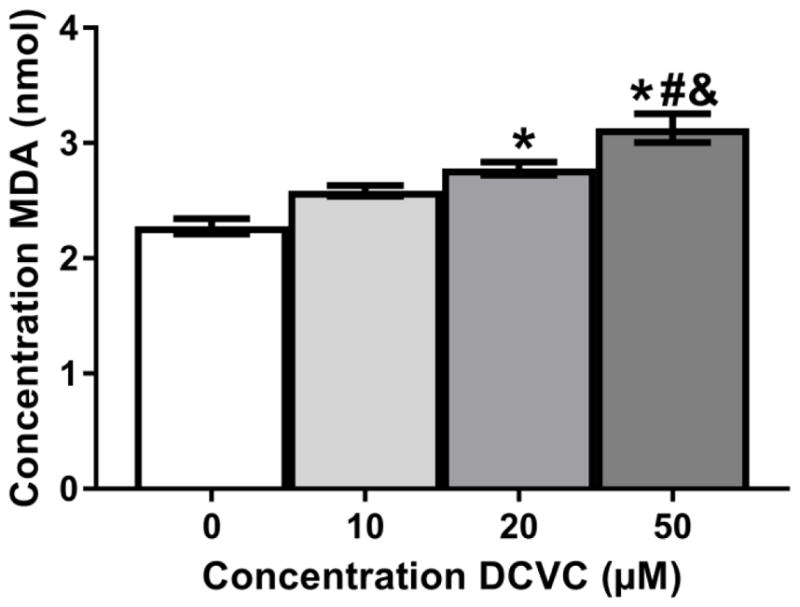
Cells were treated for 24 h with 0 (control), 10, 20, and 50 μM DCVC or 20 μM tert-butyl hydroperoxide (positive control). As a proxy measure for lipid peroxidation, malondialdehyde (MDA) concentrations were quantified using thiobarbituric acid (TBA), which generates a MDA-TBA adduct when in contact with MDA. The MDA concentrations were quantified colorimetrically (OD = 532). Bars represent means±SEM. *Significantly different compared to control (P<0.004). #Significantly different compared to 10 μM DCVC (P=0.0020). &Significantly different compared 20 μM DCVC (P= 0.0389). Data were analyzed by one -way ANOVA with posthoc Tukey multiple comparisons. N=4 independent experiments. All experiments were performed in triplicate. TBHP (20 μM) was used as a positive control (2.87 nmol +/− 0.167 nmol).