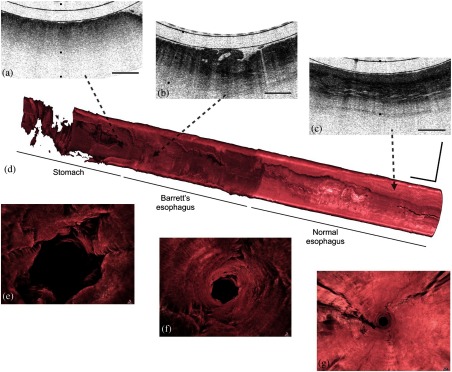
Fig. 5.
Tethered capsule endomicroscopy data from a patient with a diagnosis of Barrett’s esophagus and high-grade dysplasia, with intramucosal carcinoma. (a–c) Portion of a cross-sectional tethered capsule microscopy image of (a) the stomach, (b) Barrett’s esophagus mucosa with architectural atypia suggestive of high-grade dysplasia, and (c) squamous mucosa at the distal, mid, and proximal ends of the esophagus, respectively. (d) A three-dimensional representation of the tethered capsule endomicroscopy data showing a 4-cm segment of Barrett’s esophagus with multiple raised plaques and nodules, one of which corresponds to the features in (b). (e–g) Three-dimensional fly-through views of: (e) the stomach, (f) Barrett’s segment, and (g) squamous mucosa showing a clear difference between the superficial appearance of the rugal folds of the stomach, the crypt pattern of Barrett’s esophagus, and the smooth surface of the squamous mucosa. Tick marks and scale bars represent: (a–c) 1 mm; scale bars: (d) 1 cm. Figure reprinted with permission from Ref. 107.

