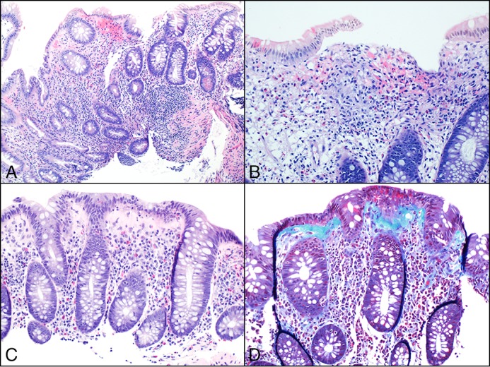
Figure 1.

Histologic images from patient 1 showing disease progression. (A) Expansion of lamina propria, architectural glandular disarray, and focal cryptitis occurring during chronic colitis with mild activity. (B) Mucosal granulomas were also identified, supporting the diagnosis of Crohn’s colitis. (C) Loss of surface mucin and intraepithelial lymphocytes, a markedly thickened collagen plate, but no evidence of glandular disarray. (D) Trichrome stain highlighting the thickened collagen under the surface epithelium, confirming the diagnosis of collagenous colitis.
