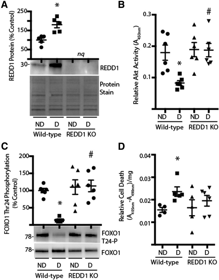
Figure 3.
REDD1 ablation prevents diabetes-induced retinal cell death. Retinas were isolated from diabetic (D) and nondiabetic (ND) wild-type and REDD1-knockout (KO) mice 4 weeks after STZ administration. A: Expression of REDD1 in retinal lysates was assessed by Western blotting. Gel loading was assessed by protein stain. B: Akt activity in retinal lysates was assessed by ELISA by using a synthetic peptide substrate. C: FOXO1 phosphorylation was assessed by Western blotting. D: Retinal lysates were assayed by ELISA for the presence of nucleosomal fragments in the cytoplasm. Values are means ± SE for two independent experiments (n = 5–7). *P < 0.05 vs. ND and #P < 0.05 vs. wild-type. Protein molecular mass in kDa is indicated at the left of blots. nq, no quantification of REDD1 expression was performed on retinal lysates from REDD1-deficient mice.

