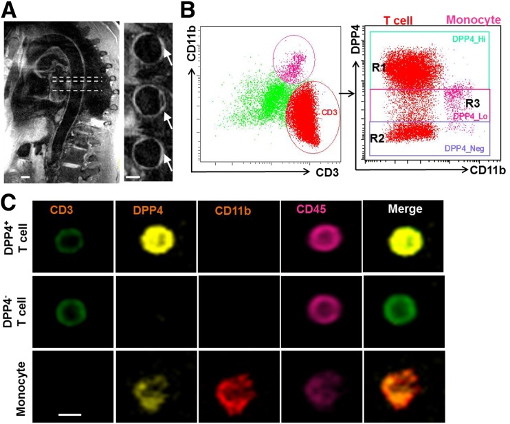
Figure 1.
A: High-resolution three-dimensional dark-blood MRI was used to confirm the aortic atherosclerotic plaque in patients with atherosclerotic disease. Arrows indicate the thickening of the aortic wall. Scale bars, 10 mm. B and C: DPP4 expression on circulating immune cells. Peripheral blood mononuclear cells were isolated from healthy volunteers, and CD11b+ monocyte and CD3+ T cells were gated for the detection of DPP4 using an imaging flow cytometer. Representative scatter plots (B) and cell images (C) are shown. Scale bar, 5 μm. R1, region 1 (region with high DPP4 expression); R2, region 2 (region without DPP4 expression); R3, region 3 (region with low DPP4 expression).

