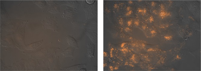
Figure 6.

Fluorescence micrographies of HeLa cells. Bright-field images are superimposed to the red emission channel after incubation with 5 μM TMR-brHis2 for 30 min at 37 °C (left) and the same experiment in the presence of 5 μM Pd(en)Cl2 (right). The complex was preincubated (1:1) in water for 10 min before the addition. The cells were washed twice with PBS before being observed in a fluorescence microscope. All incubations were made in Dulbecco’s modified Eagle medium completed with 10% fetal bovine serum. TMR: tetramethylrhodamine.
