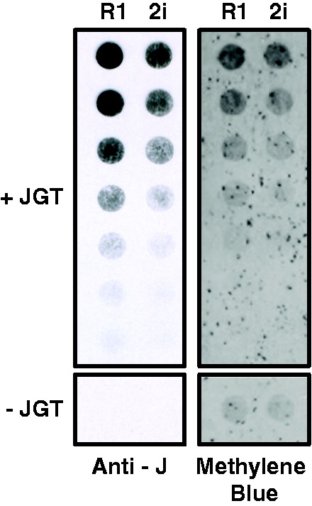
Figure 5.

Quantitation of 5hmU in mESC genomic DNA. Genomic DNA isolated from two mESC lines (R1 and 2i) was incubated with JGT and UDP-glucose, spotted onto nitrocellulose in a 2-fold dilution series and levels of glucosyl-5hmU detected by base J antisera (Anti-J). The dependence of the assay on the JGT labeling reaction is indicated below each blot by lack of signal from the highest DNA concentration assayed without the addition of JGT (−JGT). Methylene blue staining of the blot controls for DNA loading. Shown is a representative blot from three independent experiments.
