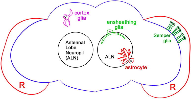
Figure 1. Glial subtypes in the adult Drosophila CNS.

Schematic diagram of the adult fly brain illustrates representative classes of glia. In the cortical regions of the CNS, cortex glia (magenta) surround neuronal cell bodies, likely providing important metabolic and functional support. Neuropil areas of the central brain, which house axonal and dendritic projections, contain two major glial subtypes: Ensheathing glia (light green) enwrap neuronal extensions and appear to serve as the primary immune responders in adult animals, while astrocytes (red) modulate synaptic signaling. Representative diagrams of ensheathing glia and astrocytes within the antennal lobe neuropil (ALN) are shown. The entire nervous system is covered by a double layer of surface glial cells (blue outline), which offers a protective barrier between the CNS and the circulating hemolyph. A more recently identified cohort of glial cells in the adult retina (R), Semper cells (dark green), regulate photoreceptor function in a manner comparable to mammalian Mueller glia.
