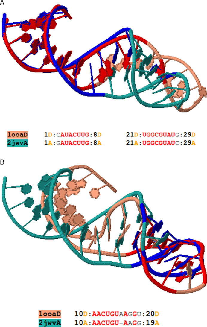
Figure 4.
(A) The first alignment of Rclick for two RNA aptamer structures, PDB codes 2JWV chain A (green) and 1OOA chain D (salmon) with SO of 62.07% and RMSD of 1.75 Å. The superimposed residues of 2JWV chain A and 1OOA chain D are shown in blue and red, respectively. The regions, spanning residues 1–8 and 21–29 are aligned with one another. With conformational change, Rclick shows the second alignment of the regions of residues 10–20 of 1OOA chain D and 2JWV chain A with SO of 31.03% and RMSD of 1.91 Å in (B).

