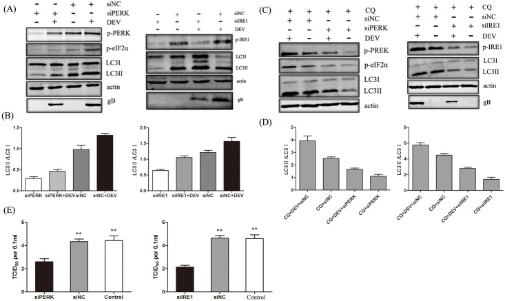
Fig 3. Effects of PERK-eIF2α and IRE1-XBP1 pathways on DEV replication.
(A) DEF cells were transfected with a Negative Control siRNA (siNC) or siRNAs directed against PERK or IRE1 (100 pmol/ml), followed by DEV infection. Whole-cell protein extracts were collected at 48 hours post-infection and then subjected to immunoblotting analysis of PREK, p-PERK, eIF2α p-eIF2α, IRE1, p-IRE1, LC3, and β-actin using the indicated antibodies. (B) Ratio data of LC3-II to LC3-I in treated DEF cells. (C) CQ was added to each sample in presented treated DEF cells.Whole-cell protein extracts were collected at 48 hours post-infection and then subjected to immunoblotting analysis of PREK, p-PERK, eIF2α p-eIF2α, IRE1, p-IRE1, LC3, and β-actin using the indicated antibodies. (D) Ratio data of LC3-II to LC3-I in treated DEF cells. (E) The virus yields were determined at 48 hours post-infection and expressed as TCID50 per 0.1 ml.

