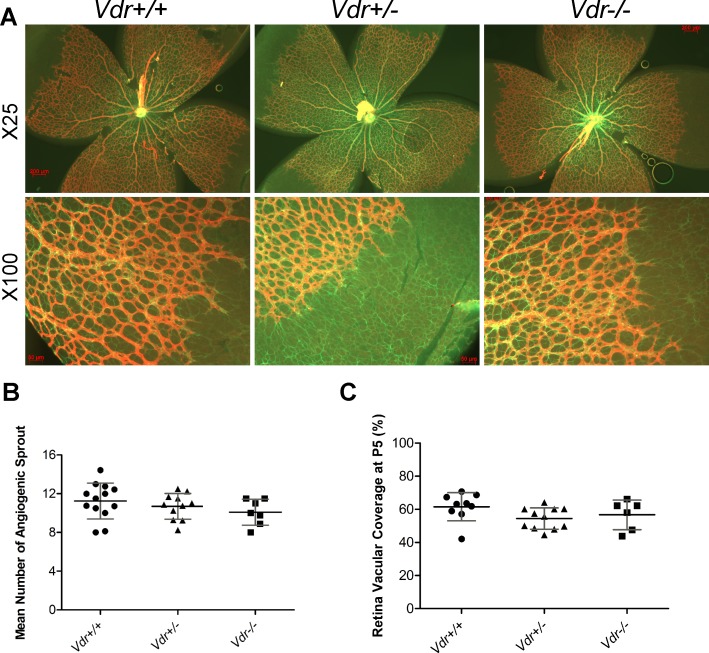
Fig 1. The development of superficial layer of retinal vasculature is independent of Vdr expression.
(A) Demonstrates GFAP and Col IV stained retinal vessels prepared from postnatal day 5 (P5) Vdr +/+ and Vdr -/- mice. Please note similar expansion of astrocytes (green, GFAP) and progression of expanding vessels (red, Col IV). Scale bar = 200 μm for x25 and Scale bar = 50 μm for x100 images. (B) The mean number of angiogenic sprouts at the angiogenic fronts were quantified per field (x100) in each retina. (C) Coverage of retinal vasculature relative to total retina area were measured for each retina and is shown as a percentage. (n≥ 5; each point represents one mouse).

