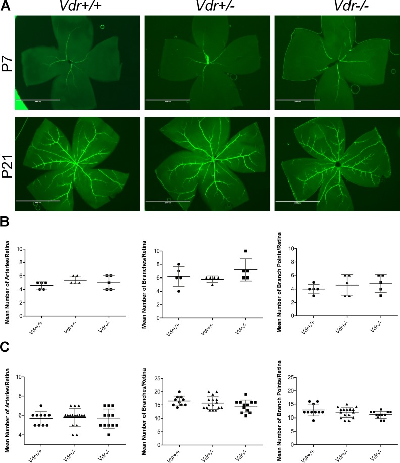
Fig 2. The organization of major blood vessels and development of primary retinal vascular plexus is not affected by Vdr-deficiency.
(A) Retinas from P7 and P21 mice were wholemount stained with anti-α-smooth muscle actin and imaged at x12.5. Mean number of major arteries, branches, and branch points were quantified per retina and shown, respectively, in (B) for P7 and (C) for P21. (n≥ 5; each point represents one mice) Scale bar = 2,000 μm.

