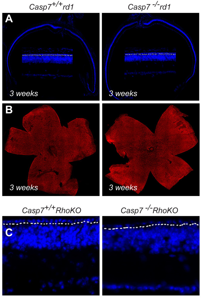
Figure 2.
Loss of Casp7 does not affect rod degeneration. A: Retinal cross section of 3-week-old Casp7+/+_rd1 and Casp7–/_–rd1 mice stained with 4´,6-diamidino-2-phenylindole (DAPI) showing no difference in the thickness of the outer nuclear layer in the central retina (n = 3 animals; the zoomed-in view shows one row of cells above the dotted line which separated the outer nuclear layer from the inner nuclear layer). B: Retinal flat mounts stained with cone arrestin (red signal) showing no difference in the distribution of cones at the onset of cone death (genotype and age same as in panel A). C: Retinal cross section of 17-week-old Casp7+/+_Rho-KO and Casp7–/–_Rho-KO mice showing no difference in the thickness of the outer nuclear layer in the central retina (n = 3 animals; the dotted line separates the outer nuclear layer from the inner nuclear layer).

