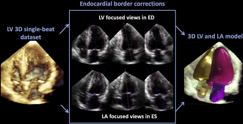
Figure 1.

Automated technique for left-heart 3D chamber quantification. Following initial fully automated detection of LV and LA endocardial surfaces from a high-frame rate single-beat 3DE data set (left), the software allows the user to perform manual corrections of the endocardial boundaries when needed (center), resulting in 3D casts of the cardiac chambers (right). The optional corrections are performed in anatomically correct nonforeshortened 2D planes showing focused long-axis views of the left ventricle (top) and left atrium (bottom), both automatically extracted from the 3D data set. (Note that the program displays right ventricular and atrial casts but no volume values are provided because they have not been validated.)
